Why is collenchyma flexible while sclerenchyma is rigid?
Week 10 – Simple Tissues: Parenchyma, Collenchyma & Sclerenchyma
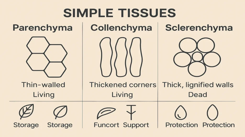
Simple tissues parenchyma, collenchyma, sclerenchyma their structure, functions, and locations, with microscope sections and micrometry practice.
Learning Outcomes
By the end of Week 10, students will be able to:
- Define simple tissues and distinguish their three major types.
- Describe the structure, function, and plant locations of parenchyma, collenchyma, and sclerenchyma.
- Identify these tissues in hand-cut sections under the microscope.
- Prepare labeled drawings showing characteristic cell features.
- Measure cell size using an eyepiece micrometer.
- Relate tissue type to mechanical support, storage, and metabolic activity.
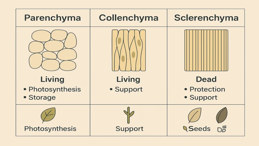
Parenchyma
Structure:
- Living, thin-walled, isodiametric cells.
- Large intercellular spaces.
- Primary wall only (cellulose + pectin).
- Prominent nucleus + vacuole.
Functions:
- Storage (starch, water, food)
- Photosynthesis (chlorenchyma)
- Aeration (aerenchyma)
- Regeneration & wound healing
Locations:
- Cortex & pith of stems/roots
- Mesophyll of leaves
- Fruit flesh
- Xylem & phloem parenchyma
Collenchyma
Structure:
- Living cells with unevenly thickened cell walls
- Thickening at corners (angular) or uniform around walls (lamellar)
- Cellulose + pectin + hemicellulose thickening
- Elongated cells
Functions:
- Mechanical support to growing organs
- Flexible support without restricting growth
Locations:
- Leaf petiole
- Young stems below epidermis
- Midrib of leaves
Sclerenchyma
Structure:
- Dead at maturity
- Very thick lignified secondary walls
- Two main types:
- Fibres (long, tapered)
- Sclereids (stone cells, branched, isodiametric)
Functions:
- Strength & rigidity
- Protection (seed coats, nutshells)
- Mechanical reinforcement in mature organs
Locations:
- Hard seed coats (testa)
- Coconut husk fibres
- Vascular bundles (fibres)
- Gritty texture of pear fruit (sclereids)
The approach followed at E Lectures reflects both academic depth and easy-to-understand explanations.
People also ask:
Collenchyma has pectin-rich, uneven primary wall thickening, allowing flexibility. Sclerenchyma has lignified secondary walls, making it rigid.
Parenchyma, because the cells remain living and retain meristematic ability.
They provide protection and contribute to the gritty texture due to thick lignified walls.
Rarely. It is mostly found in aerial young organs for mechanical support.
Observe wall thickness, presence/absence of lignin, and cell shape thin & round (parenchyma), thick corners (collenchyma), thick secondary wall (sclerenchyma).